
- 2026-Jun-04
Espen Berg
Our Intelligent Engineering solutions across products, plant and networks, combine our engineering expertise with advanced technologies to enable digital engineering & operations, develop autonomous products & platforms, and build sustainable energy and infrastructure
.png?width=774&height=812&name=Master%20final%201%20(1).png)
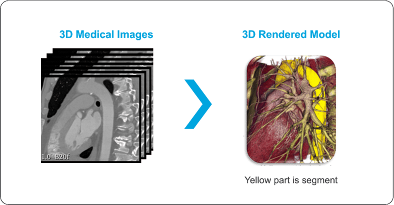
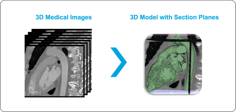
Enhanced visualization is a technique that transforms a series of 3D DICOM images from radiology data into a 3D cardiac anatomy view using an interactive application that presents different sections of the anatomy, helping find the best-fit section for desired visualization. This allows physicians to visualize and intuitively understand the cardiac anatomy for interventional studies. Different segments based on the anatomy of interest can be separated as parts and can be exported as .STL files to be used for generating the geometric coordinates to create the 3D model and for 3D printing the separated parts.
Cardiac interventions remain technically challenging owing to the anatomical complexity and heterogeneity of cardiac pathologies. As such, multi-disciplinary pre-procedural planning assisted by advanced cardiac imaging is pivotal to successful outcomes. Modern imaging techniques offer accurate 3D renderings of cardiac anatomy; however, users are required to derive a spatial understanding of complex cardiac pathologies from a 2D projection, thus generating an “imaging gap” that limits procedural planning.
Physical cardiac anatomy modeling using 3D printing has the potential to bridge this gap and is increasingly being employed in conjunction with other transformative technologies to assess the feasibility of the intervention and the creation of customized anatomical implants or prosthetics.
Such platforms have also shown value in training and patient education. Despite important limitations, the pace of innovation and synergistic integration with other technologies are likely to ensure that 3D printing assumes a central role in delivering personalized care for patients undergoing surgical interventions.
The evolution of radiology imaging has allowed surgeons to better prepare pre-procedural surgical planning. As cross-sectional imaging has rapidly advanced in the last decade, high-resolution 3D images can routinely be obtained which help visualize complex cardiac anatomy. Despite these advances in technology, the 3D image still needs to be viewed on a 2D screen. With Enhanced Visualization, we can just see the Anatomy as a 3D model, can use options to segment part by part and understand it more clearly. With different types of 3D printing technologies and materials that can be used to create subject-specific anatomical models, such as resin, plastic, metal, or even bioprinting. Depending on the purpose and the desired level of detail, some 3D printing methods like resin-based 3D printing can produce high-resolution models with realistic colors and textures. Enhanced visualization aids in converting standard 2D medical images into 3D immersive models to select and view desired areas of interest and help in discussions and planning before seeing the real parts and before starting surgery.
A major advantage of enhanced visualization is its use in additive manufacturing, where consecutive layering of two-dimensional (2D) slices is combined to form a 3D object. It allows models to be custom-made at a relatively low cost without the need for expensive molds or casts. The models are derived from 3D reconstructed images, printed, and can be used for surgical planning, such as an endovascular abdominal aortic aneurysm repair. There are a variety of current and developing uses for the treatment of vascular disease, including the creation of models for surgical planning, education and training, and vascular device and tissue engineering.
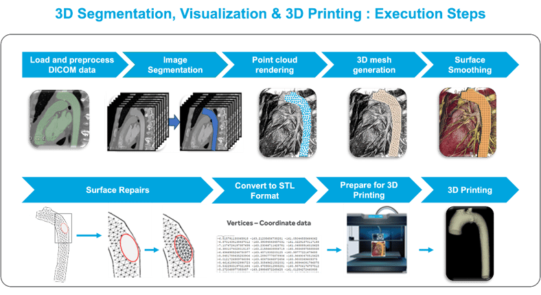
The procedure for medical enhanced visualization consists of the following steps:
Enhanced visualization opens up vast possibilities of 3D printing in the field of cardiac pathologies. Despite ongoing challenges, it offers precious insights to physicians and aids in more effective surgical/procedural planning.
About the Authors

Venkat Sudheer Naraharisetty, Lead Data Scientist
With a robust career spanning over 14 years, he has amassed extensive experience in diverse fields such as Automotive Research and Development, Computer Aided Engineering, and Data Science, with a particular focus on Artificial Intelligence. His expertise extends across a wide array of domains, including crash analysis and the development of classification models using advanced machine learning and deep learning techniques. These skills have been applied in various sectors, notably Automotive, consumer electronics and Medical Imaging.

Amol Gharpure, Senior Solution Architect – Healthcare and Life Sciences
Amol has 18+ years of embedded product development experience in the healthcare domain with valuable experience in design and development of medical devices across the product development cycle.

Srinivas Rao Kudavelly, Consultant Senior Principal - Healthcare and Life Sciences
Srinivas has over 25 years of experience which spans across Consumer Electronics, Biomedical Instrumentation and Medical Imaging. He has led research and development teams, focused on end-to-end 3D/4D quantification applications and released several "concept to research to market" solutions. He also led a cross functional team to drive applied research, product development, human factors team, clinical research, external collaboration and innovation. He has garnered diverse sets of skill sets and problem challenges. and has over 25 Patent filings and 12 Patent Grants across varied domains, mentored over 30+ student projects, been a guide for over 10+ master thesis students, peer reviewer for papers and an IEEE Senior Member (2007).
For more details on this blog, please get in touch with:
srinivasrao.kudavelly@cyient.com