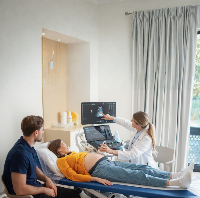
Every parent who has sat in an ultrasound room knows the moment: the technician moves the probe, an image flickers onto the screen, and for a second you see something that looks almost like your baby. Then the picture shifts, blurs, or vanishes behind a shadow, and you're back to squinting at grainy shapes and trusting the clinician to tell you everything is fine.
That gap between what parents hope to see and what an ultrasound actually shows isn't a flaw in the technology or the person holding the probe. It's physics. And it's a problem which Cyient’s Intelligent Engineering Services (IES) team has been quietly solving with a combination of clinical imaging science and artificial intelligence.
Why ultrasound can never show you "The whole baby"
Ultrasound works by sending sound waves into the body and listening for the echoes that bounce back. It's safe, affordable, and available almost everywhere, which is exactly why it has remained the backbone of prenatal care for decades. But sound waves have limits.
Bone is highly reflective, so a baby's own skull, spine, or limbs can block the beam and cast a shadow over whatever lies behind them. Tissue absorbs and weakens the signal the deeper it travels, especially in later pregnancy. The probe can only "see" from wherever it happens to be positioned, and a structure that's clear from one angle may be completely hidden from another. Add in the simple fact that babies move, and it becomes clear why even advanced 3D ultrasound machines often capture a sweep that is missing pieces of anatomy. No amount of clever rendering can fill in detail that was never captured in the first place.
"The gap between what parents hope to see and what an ultrasound shows isn't a flaw — it's physics."
A different way of filling the gaps
"Instead of treating a scan as a puzzle that's simply missing pieces, Cyient's approach treats it as a partial but reliable source of truth — one that can be intelligently completed using everything medicine already knows about how babies grow." [1]
The idea draws on two things that already exist in every prenatal clinic: the measurements a sonographer takes during a scan, such as head circumference, abdominal circumference, and femur length, and decades of population-level growth data captured in fetal growth charts, known clinically as nomograms. These charts describe, with remarkable precision, how a fetus proportions typically change week by week, and even how much natural variation exists between perfectly healthy babies at the same stage.
By combining what an ultrasound image directly shows with what medical science already knows about fetal development at that gestational age, an AI system can build a three-dimensional model of the baby that is both personal and anatomically grounded, rather than a generic textbook shape or an incomplete, shadow-riddled volume.
How it actually works
The process begins the moment a sonographer captures a standard 2D image — of the head, the abdomen, a limb, or any other structure routinely measured during a scan. An AI model first recognizes which body part it's looking at, then traces its precise boundaries, much the way a radiologist would outline an organ on a scan, but automatically and in a fraction of a second.
From that outline, the system calculates the same biometric measurements a clinician would take by hand. Those numbers are then used to select a reference 3D shape appropriate for the baby's gestational age, one built from the same nomogram data used in clinics worldwide, and to reshape it so that it reflects this specific baby's proportions rather than an average one. The head is adjusted using the measured head circumference, the torso using the abdominal measurements, the limbs using bone length, all while keeping the anatomy connected and proportionally coherent, not stitched together as isolated parts.
The result is rendered as a smooth, rotatable 3D representation, complete with shading, depth, and surface detail that makes it far easier for expectant parents and non-specialist care providers to understand than a handful of disconnected 2D images ever could.
Honesty built into the model
What makes this approach clinically credible rather than just visually impressive is its transparency about what it actually knows. Every part of the model can be traced back to one of three sources:
-
Category
- Directly observed
- Nomogram-constrained
- Model-derived
-
What it means
- Anatomy visible in the ultrasound itself — segmented skull boundaries, abdominal contours, femur endpoints.
- Expected anatomical relationships for the fetus's gestational age and growth percentile, drawn from established clinical charts.
- Surfaces not fully visible on the scan, generated by adapting the 3D shape prior — a plausible estimate, not a direct image.
That distinction matters enormously in a clinical setting. A complete-looking 3D image could otherwise create a false sense of certainty about structures that were never actually imaged.By keeping directly observed, growth-constrained, and model-derived information separate, the system gives clinicians a tool that enhances understanding without overstating what it can prove.
Training the system to handle real-world variety
Medical AI is only as good as the data behind it, and fetal ultrasound data is notoriously uneven, varying by device manufacturer, hospital, sonographer technique, and the countless ways a baby can be positioned in the womb.To make the model robust across all of that variability, Cyient's approach also includes a way to generate realistic synthetic ultrasound images, built from the same biometric measurements and anatomical outlines used elsewhere in the system.These synthetic images are then checked against real scans by a second AI model trained to identify anything that looks anatomically implausible.
This lets the system learn from rare gestational ages, unusual growth patterns, and difficult imaging conditions that might otherwise be underrepresented in real clinical datasets, ultimately making the technology more reliable for a wider range of patients and care settings.
What this could mean for prenatal care
- Standardized biometry — Automated segmentation and measurement that reduce operator-dependent variability.
- Faster assessment — Automated calculation of key measurements cuts down repetitive manual effort.
- Integrated growth visualization — Separate 2D views brought together into one coherent 3D context.
- Better communication — A model that's easier for parents and non-specialists to understand.
- Broader accessibility — Advanced visualizations for clinics without high-end volumetric imaging systems.
For many parents, a coherent, understandable 3D image is simply easier to connect with than a set of grainy 2D frames — an emotional benefit that shouldn't be underestimated in the middle of a pregnancy filled with uncertainty.
The bigger picture
The future of healthcare will not be defined solely by better devices or more data. It will be shaped by how intelligently we connect information, expertise, and human understanding.
Cyient's fetal 3D visualization innovation demonstrates how AI can extend the value of existing clinical workflows, helping clinicians gain clearer insights and enabling expectant parents to engage more meaningfully with one of life's most important moments.
As healthcare increasingly embraces AI-augmented imaging and visualization, Cyient remains focused on developing solutions that combine engineering excellence, clinical relevance, and responsible innovation, delivering technology that is not only intelligent, but impactful.
Reference:
[1] U.S. patent application, US 2026/0256457 A1, titled "System and Method of Generating an Anatomical Three-Dimensional Model," Srinivas Rao Kudavelly , Cyient Ltd, India